Neuromuscular
Muscle – Mouse nerve
Sample preparation according to the following protocol:
- Classic PFA- Glutaraldehyde, Osmium, Epon fixing.
- Ultrafine section – 1 minute UranyLess contrast – 1 minute Lead Citrate.
Photos taken with a Hitachi HT7700 Transmission Electron Microscope by Nacer BENMERADI (R&D – Delta Microscopies – France).
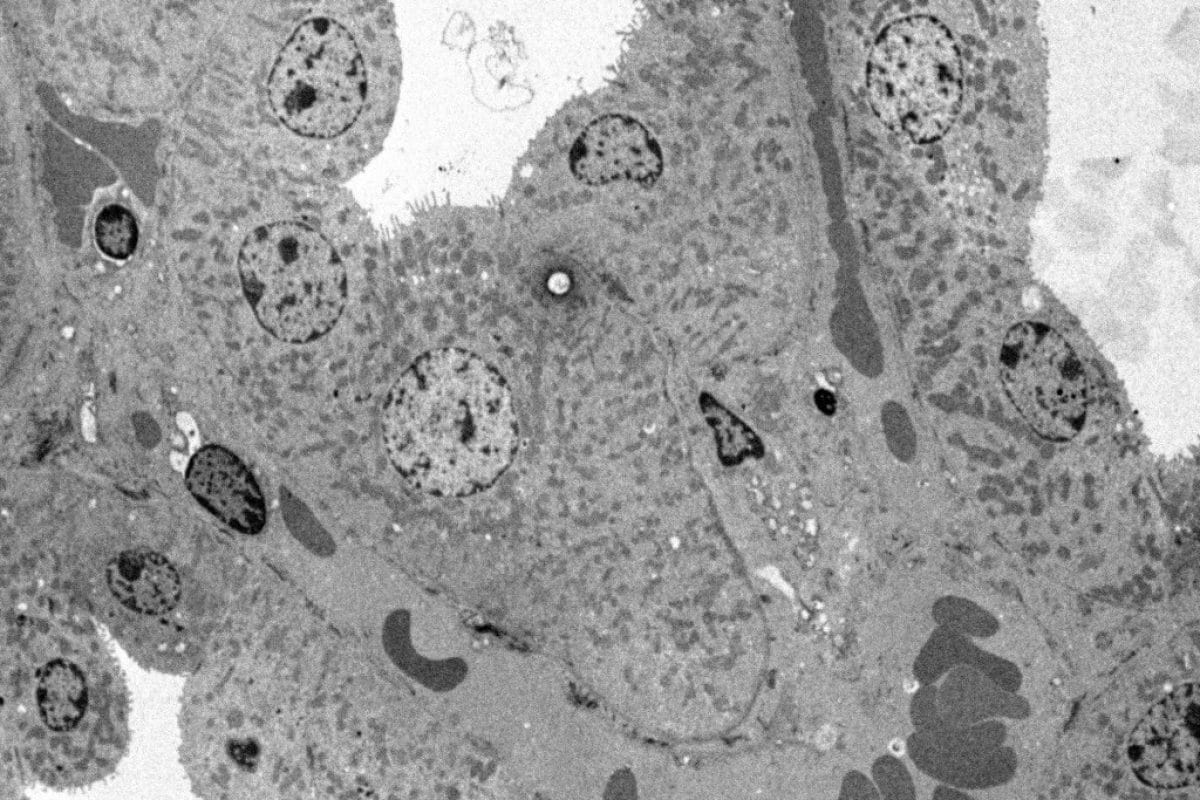
Kidney
Mouse Kidney
Sample preparation according to the following protocol:
- Classic PFA- Glutaraldehyde, Osmium, Epon fixing.
- Ultrafine section – 1 minute UranyLess contrast – 1 minute Lead Citrate.
Photos taken with a Hitachi HT7700 Transmission Electron Microscope by Nacer BENMERADI (R&D – Delta Microscopies – France).

Andrenal Gland
Adrenal gland Sahara gerbil
Sample preparation according to the following protocol:
- Classic PFA- Glutaraldehyde, Osmium, Epon fixing.
- Ultrafine section – UranyLess contrast – Lead Citrate.
Photos taken with a Hitachi HT7700 Transmission Electron Microscope by Nacer BENMERADI (R&D – Delta Microscopies – France).

Ovarian - Ovarian Follicle - Uranyless
Mouse ovarian follicle
We have tested UranyLess on different accessories cells constituting the mouse ovarian follicle.
Sample preparation according to the following protocol:
- Classic PFA- Glutaraldehyde, Osmium, Epon fixing.
- Ultrafine section – UranyLess contrast – Lead Citrate.
Photos taken with a Hitachi HT7700 Transmission Electron Microscope by Nacer BENMERADI (R&D – Delta Microscopies – France).
Navigation
Contact
22b route de Saint Ybars 31190
Mauressac France
+(33) 5 61 73 60 14
info@deltamicroscopies.com


