customer results
Yeast
Preparation of the sample according to the following protocol:
- Standard fixation with Glutaraldehyde – Osmium – Embedding in Epon.
- Contrast with UranyLess followed by Lead Citrate.
Here are some results from Jeannine Lherminier (INRA – Dijon).
Photographs taken using Transmission Electron Microscopy
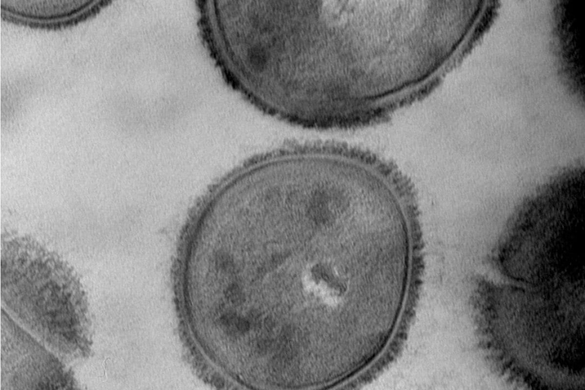
Bacteria
Bacteria in cross-section Here are the results from Christine Longin (INRA Jouy en Josas).
Sample preparation according to the following protocol:
- Standard fixation with glutaraldehyde, osmium, and embedding in Epon
- Ultrathin sectioning, double contrast with Uranyless followed by lead citrate
Photographs taken using Transmission Electron Microscopy
Polymersomes-Uranyless
The IMRCP Laboratory in Toulouse, led by Anne-Françoise Mingotaud, tested UranyLess in comparison to uranyl acetate, which is at an acidic pH of 4 (seems to disrupt the molecular structure organization). They also compared observations using Cryo-SEM (scanning electron microscopy).
Negative Staining in Uranyl Acetate pH 4
Plant Tissu
Trematodes
We present some results from Yann Quilichini (Microscopy Platform at the University of Corsica – Corte).
Sample preparation according to the following protocol:
- Standard fixation with glutaraldehyde, osmium, embedding in Spurr resin.
- Thin sections – contrast with aqueous UranyLess followed by lead citrate (according to Reynolds).
Photographs taken using Transmission Electron Microscopy.
Acculina Crustaceans (small parasitic crustacean)
Sacculina Crustaceans (small parasitic crustacean)
We present some results from Djédiat Chakib (Muséum National d’Histoire Naturelle, Paris).
Sample preparation according to the following protocol:
- Standard fixation with glutaraldehyde, osmium, embedding in epoxy.
- Thin sections – contrast with aqueous UranyLess at 60°C on a heating plate without lead citrate in post-staining.
Photographs taken using Transmission Electron Microscopy.
Reconstructed Epidermis
We present the results from Audrey Houcine (CMEAB Toulouse).
Sample preparation according to the following protocol:
- Standard fixation with glutaraldehyde, osmium, Epon/Araldite
- Ultrathin sectioning, double contrast with Uranyless followed by lead citrate
Photographs taken using Hitachi HT7700 Transmission Electron Microscopy by Audrey Houcine.
Automated Contrast with LEICA EM Stain - Uranyless
Chantal Cazevieille from CRIC/IURC, INSERM Montpellier, tested aqueous Uranyless in the Leica grid contrast automaton on various tissues, including drosophila, atrium of the heart, retina, cochlea, and ileum (digestive tract). The tissues were fixed according to the standard protocol with 2.5% glutaraldehyde in PHEM buffer, followed by post-fixation in 0.5% osmium and 0.8% potassium ferrocyanide for 2 hours at room temperature. Sections were collected on single-hole or 200-mesh grids.
The grids were treated with Uranyless for 7 minutes, followed by lead citrate for 7 minutes.
We present here only a few images taken with the Hitachi Transmission Electron Microscope using an AMT digital camera.
You will note that the combined action of potassium ferrocyanide and Uranyless reveals the cyto-membranes in the ileum more prominently.
Navigation
Contact
22b route de Saint Ybars 31190
Mauressac France
+(33) 5 61 73 60 14
info@deltamicroscopies.com


